Causes of a soft forehead in a child. Intracranial pressure in infants: treatment for symptoms. Homework for parents
After all, the first month of a child’s life becomes for him the first critical period after birth: it is characterized by the intense work of all organs and systems of the body “responsible” for the adaptation of the newborn to environmental conditions that are fundamentally new for him. By the end of this period, all transition processes should be completed, however, under the influence of unfavorable environmental conditions, with aggravated pregnancy and childbirth, the natural adaptation processes for a newborn can take on a pathological direction and lead to a neurological disease of the child.
It is at this time that it is necessary to visit a neurologist for the first time - usually just to make sure that everything is fine with the baby; but if this is not the case, in order to identify and “capture” the pathology at the very beginning, to prevent the disease from developing. To determine the level of development of the child and exclude neurological pathology, it is important not only to assess the formed reactions to light, sound, motor and psycho-emotional activity of the newborn, but also his appearance (in fact, it is this last topic that my article will mainly be devoted to).
So, what will a neurologist first of all pay attention to when examining a one-month-old baby? On the shape and size of his skull, facial expression, posture, appearance of the skin. Why is this so important? Why are our worries and worries often associated with the presence of deviations in the child’s appearance, especially if this is a change in the shape and size of the skull? This is primarily due to the fact that such changes can be a diagnostic sign of serious diseases - hydrocephalus and microcephaly.
The shape and size of the skull is a possible pathology
Hydrocephalus - this is an excessive increase in the size of the skull and fontanelles, caused by an increase in the amount of cerebrospinal fluid in the cranial cavity. With this disease, the shape of the skull also changes - its cerebral part significantly predominates over the facial part, the frontal part protrudes sharply forward, and a pronounced venous network is observed in the area of the temples and forehead.
Microcephaly - this is a reduction in the size of the skull and early closure of the fontanelles. With congenital microcephaly, the size of the skull is small from birth, the cranial sutures are narrowed, the fontanelles are either closed or small in size. Subsequently, a slow rate of growth in head circumference is noted, so that sometimes a 2-3 year old child’s skull size is almost the same as at birth. With microcephaly, the skull has a specific shape: the cerebral part of the skull is smaller than the facial part, the forehead is small, sloping, the line of the forehead and nose is sloping.
Conditions such as hydro- and microcephaly subsequently lead to delayed mental and physical development and therefore require correction from a very early age!
...or a reason for further examination?
But should every deviation from the norm clearly indicate a pathological condition? Of course not! Clinical observations show that there are many factors that influence the shape and size of the head. Of course, even a slight increase or decrease in the circumference of the skull in a newborn compared to the age norm can be considered a risk factor for the development of hydrocephalus or microcephaly, but you should not panic when you discover that the baby’s head is slightly larger or smaller than normal: this circumstance should first of all, become a signal for the need for additional examinations to exclude pathological conditions. What kind of examinations are these?
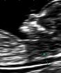
- An absolutely safe and reliable method is neurosonography(ultrasound examination of the brain through the large fontanel). This study will help not only to see changes in the structure of the brain and signs of increased intracranial pressure, but also to evaluate blood flow through the main vessels of the brain.
- An even more reliable method is nuclear magnetic resonance of the brain (NMR), but this study for children is carried out under general anesthesia, so it is carried out only for sufficiently compelling indications.
- In this case, consultations with an ophthalmologist and a neurosurgeon are also necessary.
"Homework" for parents
In addition, right from birth you can independently control increase in baby's head circumference, which is one of the main indicators of normality and pathology. How to do this correctly?
- Measure the child's head circumference weekly and record the resulting numbers in a specially kept notebook.
- When measuring, place the measuring tape at the most protruding points of the skull (frontal and occipital protuberances).
- To avoid misunderstandings, the measurement must be carried out by the same person.
In addition to the increase in head circumference, you can control increase in chest circumference, which is one of the general anthropometric indicators of child development. For this:
- Measure your chest circumference weekly on the same day you measure your head circumference;
- Place the measuring tape at the level of the baby's nipple line.
Why is such “amateur activity” needed? By taking these simple measurements, you will help the doctor draw up an objective picture of the child’s development, and you yourself can have peace of mind, excluding the possibility of developing serious diseases (normally, the monthly increase in head circumference in the first three months of a full-term baby should not exceed 2 cm per month; up to a year, the circumference The chest is approximately 1 cm larger than the child’s head circumference).
Well, now a few words about what can and should be normal and what is pathological. I tried to frame the conversation on this topic in the form of answers to questions that most often concern young parents.
What determines the shape of a newborn’s skull?
Normally, as a child passes through the birth canal, the bones of the skull overlap each other. Features of the course of the birth process affect changes in the shape of the skull. In the event of a complicated birth, a sharp juxtaposition of the skull bones may occur on top of each other, and this will lead to its deformation, which will persist for quite a long time.
A change in the shape of the skull can be expressed in the preservation swelling soft tissues of the head in the place where the child moved forward along the birth canal. The swelling disappears within the first 2-3 days. Cephalohematoma(hemorrhage under the periosteum) also changes the shape of the skull. It resolves more slowly than swelling, and this process requires the supervision of specialists (neurologist, surgeon).
Changes in the shape of the skull are also associated with age-related characteristics. In a newborn, the skull is elongated in the anteroposterior direction, and after a few months the transverse size of the skull will increase and its shape will change.
Some change in the shape and size of the skull can also occur during normal development in premature babies, or when the child is often placed on the same side, or when the child lies on his back for a long time.
How does a newborn's head grow?
The average head circumference of a newborn is 35.5 cm (the range of 33.0-37.5 cm is considered normal). The most intensive increase in head circumference in full-term babies is observed in the first 3 months - on average, 1.5 cm for each month. Then the growth decreases slightly, and by the age of one year the child’s head circumference is on average 46.6 cm (normal limits are 44.9 - 48.9 cm).
The head circumference of a premature baby increases faster than that of a full-term baby, and the increase is most pronounced during the period of active weight gain, and by the end of the 1st year of life it reaches normal values. The exception is very premature babies.
However, one should always keep in mind that even with normal development of a child, there may be physiological deviations from average values, which are often associated with constitutional characteristics or environmental influences.
What is a fontanel in a child?
The fontanelles are located in the area where the bones of the skull meet. Front, big , the fontanel is located between the frontal and parietal bones. At birth, it measures from 2.5 to 3.5 cm, then gradually decreases by 6 months and closes at 8-16 months. Rear, small , the fontanel is located between the parietal and occipital bones. It is small in size and closes by 2-3 months of life.
In pathological processes accompanied by increased intracranial pressure, the fontanelles close later, and sometimes they open again. Small sizes of the anterior fontanel may be a variant of the norm if they are not accompanied by a decrease in the circumference of the skull, the rate of its growth and a delay in psychomotor development.
The above signs do not limit the variety of possible deviations in a young child. However, it should be borne in mind that any unusual appearance of a child requires a thorough examination and monitoring of his growth and development.
When and how should a neurologist examine a child?
The development of a young child is a very sensitive sign of the state of the body. It depends both on hereditary characteristics and on a complex set of social conditions and requires dynamic monitoring by doctors. Don’t forget to show your baby to specialists within the prescribed time frame - 1, 3, 6, 12 months!
If you invite a specialist to your home, you must consider the following:
- the examination of the child should be carried out on a changing table or other soft, but not sagging surface;
- the environment should be calm, eliminate distractions if possible;
- It is advisable to carry out the examination 1.5-2 hours after feeding;
- the air temperature in the room should be about 25° C, the lighting should be bright, but not irritating.
In conclusion of the article, I would like to remind you once again: do not delay your visit to a neurologist, remember - the timeliness of all health-improving, preventive and therapeutic measures aimed at ensuring its normal development depends on the correct assessment of the newborn’s health, and only a specialist can give a correct assessment!
A small dimple on the crown of the child - the fontanelle - performs an important task during the birth of the baby. And even after birth, she is assigned a serious role, and along with this, special attention from mothers and doctors.
Shape and size of a newborn's head
The head shape of newborn babies can be not only round, but also elongated, flattened, ovoid - and all these options are considered the norm. Why is this happening?
By the time they are born, the skull bones of babies are not yet very dense (they will have to completely harden during the first year of life), and the seams between them have not yet had time to heal. During birth, the bones overlap each other, allowing the baby to move out more easily. That is why, after a natural birth, the shape of the head is, as a rule, slightly elongated, while in small “Caesareans” it is smooth and round. Due to the vicissitudes of traveling through the birth canal, a baby may be born with an asymmetrical head, and sometimes also with a lump (cephalohematoma) or edema (the so-called birth edema).
At birth, the baby's head is approximately 2 cm larger in circumference than the chest. But it happens that these sizes increase even more: this happens if cerebrospinal fluid accumulates in the cranial cavity. Then the upper part becomes larger than the lower part, a heavy forehead hangs over the eyes and nose, and doctors talk about hydrocephalus. This problem can arise if during pregnancy a woman suffered a severe infection that affected the unborn baby. In this case, doctors will immediately begin treatment of the child, and in a few months his head may approach normal size.
The situation is considered more serious when the newborn, on the contrary, has a too small head (microcephaly). Sometimes this happens due to genetic disorders that will prevent the baby from developing normally. Fortunately, in many cases the reason for the unusual shape or size of the head turns out to be much simpler: the child can inherit all these features from his parents.
Only a doctor can correctly assess the baby’s head circumference, so there is no point in parents arming themselves with a centimeter themselves. But this indicator will tell specialists whether the child’s brain is developing correctly.
Normally, newborns have a head circumference of 34-36 cm. At first, the head grows quite quickly, by about 1.5 cm per month; after 3 months - by 0.5-1 cm and by 6 months it reaches 43 cm in girth. If the baby is far ahead of or behind the norm, this may indicate problems.
Comment on the article "Newborn head: shape, size, fontanelle. Is everything okay?"
Section: -- gatherings (Shape and size of the child's head). Newborn head: shape, size, fontanel. OH in a child at 32-34 weeks. Newborn head: shape, size, fontanel. Everything is fine? When will the fontanel overgrow and what should the circumference be...
Head shape. Independent medical examination of children. Adoption. Discussion of adoption issues, forms of placing children in families. Everyone is shocked, since the child has a head like a natural pumpkin. The neurologist is ready to write a diagnosis, the child was prescribed a GM ultrasound and EEG...
Newborn head: shape, size, fontanel. Everything is fine? When will the fontanelle close and what should the child’s head circumference be? Print version. What can the shape of a newborn’s head and its size tell parents?
What are the sizes of your fontanelle and chest/head? I don’t know how you can measure the fontanel diagonally, but if it’s on both sides, then it turns out. To my shame, I don’t know: (We don’t bother with circles, the doctor tells us that everything is fine, the fontanel is in good shape (that’s it. ..
Fontana and head size. Age standards. A child from birth to one year. Six fontanelles of an infant. The fontanel of a newborn: the size of the main fontanel. The fontanel in an infant. Newborn head: shape, size, fontanel. Everything is fine?
About the shape of the head. Medical issues. A child from birth to one year. Mommies, please tell me, at what age does a child’s head shape develop? My son was born with a slightly elongated head shape and a flat back of the head, but this, in my opinion, is not...
Newborn head: shape, size, fontanel. Everything is fine? Normally, it should neither swell nor sink; touching the fontanel with your fingers, you can easily feel the pulsation. Each baby has its own rate of overgrowth of a large fontanel - this is normal...
Newborn head: shape, size, fontanel. Everything is fine? Normally, it should neither swell nor sink; touching the fontanel with your fingers, you can easily feel the pulsation. Eh, how can we fix the back of the head and make it even? head? This is a really annoying question...
Sunken fontanel! Medical issues. A child from birth to one year. Care and education of a child up to one year: nutrition, illness, development. Newborn head: shape, size, fontanel. Everything is fine?
Newborn head: shape, size, fontanel. Everything is fine? Normally, newborns have a head circumference of 34–36 cm. At first, the head grows quite quickly, by about 1.5 cm per month; after From the moment of birth, the baby’s body quickly...
Head shape. Is the head aligned? Maybe someone had children like this - how is the dynamics after 1 year, 2 years? Does the size of the head depend on the weight of the child or how to defeat cockroaches. But it turns out that my head has grown by 14.5 cm in a year.
Head size %). ...I find it difficult to choose a section. Child from 1 to 3. Raising a child from one to three years: hardening and development, dear mothers! Does anyone know what the approximate head size is for a 3 year old child? I really need it! help me please! :) thanks in advance.
head volume (survey). Medical issues. Child from 1 to 3. Raising a child from one to three years: hardening and development, nutrition and head volume (survey). in my life I have never bothered with the problems of any additional volumes, I have never measured anything for a child, except...
Big head. Age standards. A child from birth to one year. The opinions of doctors were different, I listened to a neurologist from the Institute of Pediatrics, who argued that the child had a compensated form and nothing needed to be done.
A big head is a sign of what? for some reason no one writes the size of the head, at 3 years old we have 53.5 cm (by the way, at 4 years old 54), my mother-in-law says that IMHO it’s not so much about the shape of the head, but about how it affects the development of the child. Well, in general, it’s up to you to decide whether it’s worth it.
Diagnosis based on the size of the newborn. - gatherings. Other children. Diagnosis based on the size of the newborn. Girls who have encountered a similar situation, the child has a head size of 31.5 cm and a chest size of 32 cm. Based on this, a very bad diagnosis is made.
Newborn head: shape, size, fontanel. Everything is fine? A small dimple on the crown of the child - the fontanelle - performs an important task during the birth of the baby. The situation is considered more serious when the newborn, on the contrary, has too small...
Newborn head: shape, size, fontanel. Everything is fine? The fontanel of a newborn: the size of the child’s main fontanel by month and the main fears of parents. 6–7 months.
Newborn head: shape, size, fontanel. Everything is fine? A small dimple on the crown of the child - the fontanelle - performs an important task during the birth of the baby. The only other thing I can say is the shape of the head with a very convex nape in both...
Head dimensions.. Medical issues. A child from birth to one year. Girls, tell me, what is the head circumference of your children at 3 months? Ours is now 38-40 cm. I’m wondering if we buy size 44 hats, won’t they hang on our heads later?
The last time we went to see a neurologist, she said that everything was fine with us, and there was nothing to worry about, everything was fine, the child was healthy, the reaction to all vaccinations was normal. From birth we have a slightly large head; I myself am thin. The question is: I recently noticed, or so it seems to me, that she has a very pronounced convex forehead. We were told that this happens with ICP. But my daughter sleeps well, her appetite is normal, she plays, sings, dances, well, in a word, I don’t see anything wrong, but sometimes she gets nervous and capricious. Why is the forehead growing, do we need treatment again? I’m very worried, we shaved her head, maybe that’s why it seems so to me.
mirishka 12 Jul 2010
Aimka 12 Jul 2010
Are you taking any vitamins now?
mirishka 12 Jul 2010
Aimka 12 Jul 2010
I’m looking at a photo of your daughter here, it looks like her head isn’t that big, ours seems bigger..
Eralieva-Lyazzat July 12, 2010
We also took Kinder Biovital gel, vitamin D3, Calcium Kal, and now our daughter is stocking up on natural vitamins. My daughter lives with her grandparents in the fresh air in Chilika, because... I’m at work, according to them, everything is tttttt fine, I think in any case, I need to show the doctor my head, so that later I can calm down.. otherwise I feel anxious in my soul
Aimka 12 Jul 2010
And if the head (head circumference size) grows in accordance with age standards, then everything is in order. How correctly mirishka says that if your daughter sleeps peacefully and develops according to her age, then there is no need to torment yourself with unnecessary anxiety. Moreover, tomography and ultrasound did not reveal any signs of hydrocephalus - and this is one of the main reasons for the large volume of the head. Good luck!
Intracranial pressure in infants - signs and symptoms. How to determine increased ICP in newborns
Increased intracranial pressure is a complex disease that is difficult to treat and leads to many unpleasant consequences. This disease is especially dangerous and difficult to diagnose in infants, because they cannot complain of feeling unwell.
What is ICP in a child?
Intracranial pressure occurs due to too much (hypertension) or too little (hypotension) cerebrospinal fluid, which protects brain tissue from damage. It's called cerebrospinal fluid. Often this problem occurs due to prolonged oxygen starvation of brain cells. Intracranial pressure in a newborn that is slightly elevated is normal. After some time, as a rule, it normalizes without intervention.
Congenital intracranial pressure
There are two types of ICP: congenital and acquired. Congenital intracranial pressure in infants, which is more difficult to treat, is a consequence of birth injuries and complications during pregnancy. It is not possible to say in advance whether the baby is at risk of having this disease. During examinations, there may be no prerequisites for ICP, but according to general statistics, every fifth child experiences such a pathology. Acquired intracranial pressure in an infant occurs as a result of encephalitis, meningitis or trauma.
Note!
The fungus won't bother you anymore! Elena Malysheva tells in detail.
Elena Malysheva - How to lose weight without doing anything!
Signs of ICP in a baby
Every mother dreams of a healthy child, so it is important to be able to prevent the onset of the disease and notice its signs in a timely manner, because difficulty in the outflow of cerebrospinal fluid can cause a lot of inconvenience and pain for the newborn. Many new parents rejoice at the activity of their child, are touched when the baby arches or shakes his head, and do not think that these may be the first alarm bells.
Symptoms of intracranial pressure in infants:
- frequent awakenings at night;
- hyperactivity, increased excitability;
- premature breast refusal;
- excessive regurgitation, vomiting;
- involuntary movements of the eyeball;
- tremor;
- frequent crying for no reason;
- head rotation;
- strong reaction to changes in weather;
- lethargy;
- retardation in physical, psycho-emotional development;
- tilting the head back.
Veins on the head of a baby
Young mothers often get scared and complain to the doctor that veins are visible on the baby’s head. There is nothing wrong with this phenomenon, because the skin of a newborn is thinner than that of any adult, and the layer of subcutaneous fat is not yet sufficiently developed. Over time, the venous network will become less noticeable. In some cases, the veins swell and swell, which may be a sign of poor outflow of cerebrospinal fluid: you need to contact a neurologist as soon as possible so that he can prescribe an examination and the necessary tests.
A child has a large forehead
Sometimes the first sign of the presence of ICP is a high, convex forehead in an infant, which is characterized by some overhang of the skull at the back of the head. It is often confused with dropsy. If you notice a similar deviation, look at photos of children with this diagnosis and bring the violation to the attention of the pediatrician during the examination. This may be a sign of other diseases such as hydrocephalus or rickets. In any case, do not panic, but ask for an additional examination of the baby to make sure there is no danger.
Dehiscence of the sutures of the skull in an infant
A special feature of the newborn’s skull is the mobility of the bone plates. This is necessary to make it easier for the baby to pass through the birth canal. Sometimes a divergence of the cranial sutures in infants may occur, which returns to normal after a few months, and the fontanel heals. If this does not happen, be sure to consult with the pediatrician observing the child. He must conduct a study of the structure of the head, assess the size of the gaps between the plates and prescribe the necessary preventive measures or treatment.
Causes of intracranial pressure in children
Intracranial pressure in children under one year of age can cause many difficulties and health problems in older age. The success of treatment depends, first of all, on the timeliness of assistance provided. To identify ICP in a child, it is important to carefully observe his behavior, especially in the first 2-3 weeks of life. Sometimes it is very difficult to notice the first signs of illness.
Causes of intracranial pressure in newborns:
- hypoxia (oxygen starvation caused by entanglement of the umbilical cord or other problems);
- severe toxicosis throughout pregnancy;
- placental abruption or rapid maturation;
- difficult childbirth, birth injuries;
- careless use of medications during pregnancy;
- heredity;
- brain tumors;
- hemorrhage into the cranial cavity;
- serious birth injuries.
How does intracranial pressure manifest in infants?
Increased intracranial pressure in a child is manifested by severe anxiety, sudden mood swings and hyperactivity. If your baby often cries for no reason, think about it: perhaps this is one of the symptoms of ICP associated with headaches due to increased pressure. In addition, the baby may refuse the breast, burp frequently and profusely, turn his head and roll his eyes.
Sometimes the pressure rises temporarily, then returns to normal, so the discomfort is difficult to notice. In this case, the main symptom remains crying for no apparent reason and restless behavior, which is often attributed to colic and other problems of infancy. Remember that normally, babies under 2 months should spend most of their time asleep, crying only when experiencing discomfort due to a wet diaper or hunger. If your child wakes up more than 3 times a night, constantly cries and arches, this is a serious reason to visit the pediatrician.
How to determine intracranial pressure in a baby
Correct diagnosis of intracranial pressure in children begins with a visual examination and measurement of indicators such as head volume and the size of the fontanel: in a one-year-old child it should be completely fused. Another important point in the examination is checking the muscle tone and reaction of the baby. In 99% of cases, these methods help to notice deviations in indicators in time and recognize the violation. For the purpose of an additional safety measure, almost every child is prescribed an ultrasound of brain tissue through the fontanel opening, and in some cases an encephalogram or tomography.
How to treat intracranial pressure in infants
Remember: treatment of intracranial pressure in children is prescribed by a neurologist only after a special ultrasound examination or tomography; symptoms alone are not enough to take medications. Only after making sure that the diagnosis is correct, children are prescribed Actovegin injections, and older children are given Glycine tablets. They improve the absorption of glucose by brain cells, and also normalize metabolism and have a positive effect on sleep.
Often the cause of ICP is hypoxia (lack of oxygen). In this case, special water procedures and sedatives are prescribed as treatment. This helps improve blood circulation and oxygen saturation of the brain. As a rule, blood pressure decreases after completing a course of such treatment. Otherwise, stronger medications are prescribed.
The specialist must register the child and set a return date for a re-examination. It is often prescribed after undergoing an ophthalmologist, who must conduct an examination of the fundus, and a course of baby massage, which is necessary for the general improvement of the baby’s condition. After all the described procedures, a re-measurement of head circumference, ultrasound and visual examination are carried out. If, as a result of the examination, the doctor removes the diagnosis, your child will be registered for some time with a mandatory examination every six months.
In rare cases, an increase in the volume and accumulation of cerebrospinal fluid in the brain tissue can be serious and require surgical intervention. The operation is performed under general anesthesia; a certain amount of excess cerebrospinal fluid is removed to normalize the pressure. Postoperative rehabilitation involves taking auxiliary medications and constant monitoring by a doctor.
Video: Komarovsky on intracranial pressure
The information presented in the article is for informational purposes only. The materials in the article do not encourage self-treatment. Only a qualified doctor can make a diagnosis and make recommendations for treatment based on the individual characteristics of a particular patient.
A few words about normality and pathology. Baby at an appointment with a neurologist: the shape and size of a newborn’s skull
Your baby will soon be or has already turned 1 month old! One of the most difficult periods in a newborn’s life is behind us.
Inna Sharkova
"Guta-Clinic", Moscow, pediatric neurologist
After all, the first month of a child’s life becomes for him the first critical period after birth: it is characterized by the intense work of all organs and systems of the body “responsible” for the adaptation of the newborn to environmental conditions that are fundamentally new for him. By the end of this period, all transition processes should be completed, however, under the influence of unfavorable environmental conditions, with aggravated pregnancy and childbirth, the natural adaptation processes for a newborn can take on a pathological direction and lead to a neurological disease of the child.
It is at this time that it is necessary to visit a neurologist for the first time - usually just to make sure that everything is fine with the baby; but if this is not the case, in order to identify and “capture” the pathology at the very beginning, to prevent the disease from developing. To determine the level of development of the child and exclude neurological pathology, it is important not only to assess the formed reactions to light, sound, motor and psycho-emotional activity of the newborn, but also his appearance (in fact, it is this last topic that my article will mainly be devoted to).
So, what will a neurologist first of all pay attention to when examining a one-month-old baby? On the shape and size of his skull, facial expression, posture, appearance of the skin. Why is this so important? Why are our worries and worries often associated with the presence of deviations in the child’s appearance, especially if this is a change in the shape and size of the skull? This is primarily due to the fact that such changes can be a diagnostic sign of serious diseases - hydrocephalus and microcephaly.
The shape and size of the skull is a possible pathology
Hydrocephalus- this is an excessive increase in the size of the skull and fontanelles, caused by an increase in the amount of cerebrospinal fluid in the cranial cavity. With this disease, the shape of the skull also changes - its cerebral part significantly predominates over the facial part, the frontal part protrudes sharply forward, and a pronounced venous network is observed in the area of the temples and forehead.
Microcephaly- this is a reduction in the size of the skull and early closure of the fontanelles. With congenital microcephaly, the size of the skull is small from birth, the cranial sutures are narrowed, the fontanelles are either closed or small in size. Subsequently, a slow rate of growth in head circumference is noted, so that sometimes a 2-3 year old child’s skull size is almost the same as at birth. With microcephaly, the skull has a specific shape: the cerebral part of the skull is smaller than the facial part, the forehead is small, sloping, the line of the forehead and nose is sloping.
Conditions such as hydro- and microcephaly subsequently lead to delayed mental and physical development and therefore require correction from a very early age!
. or a reason for further examinations?
But should every deviation from the norm clearly indicate a pathological condition? Of course not! Clinical observations show that there are many factors that influence the shape and size of the head. Of course, even a slight increase or decrease in the circumference of the skull in a newborn compared to the age norm can be considered a risk factor for the development of hydrocephalus or microcephaly, but you should not panic when you discover that the baby’s head is slightly larger or smaller than normal: this circumstance should first of all, become a signal for the need for additional examinations to exclude pathological conditions. What kind of examinations are these?
- An absolutely safe and reliable method is neurosonography(ultrasound examination of the brain through the large fontanel). This study will help not only to see changes in the structure of the brain and signs of increased intracranial pressure, but also to evaluate blood flow through the main vessels of the brain.
- An even more reliable method is nuclear magnetic resonance of the brain (NMR), but this study for children is carried out under general anesthesia, so it is carried out only for sufficiently compelling indications.
- In this case, consultations with an ophthalmologist and a neurosurgeon are also necessary.
Homework for parents
In addition, right from birth you can independently control increase in baby's head circumference, which is one of the main indicators of normality and pathology. How to do this correctly?
- Measure the child's head circumference weekly and record the resulting numbers in a specially kept notebook.
- When measuring, place the measuring tape at the most protruding points of the skull (frontal and occipital protuberances).
- To avoid misunderstandings, the measurement must be carried out by the same person.
In addition to the increase in head circumference, you can control increase in chest circumference, which is one of the general anthropometric indicators of child development. For this:
- Measure your chest circumference weekly on the same day you measure your head circumference;
- Place the measuring tape at the level of the baby's nipple line.
Why is such “amateur activity” needed? By taking these simple measurements, you will help the doctor draw up an objective picture of the child’s development, and you yourself can have peace of mind, excluding the possibility of developing serious diseases (normally, the monthly increase in head circumference in the first three months of a full-term baby should not exceed 2 cm per month; up to a year, the circumference The chest is approximately 1 cm larger than the child’s head circumference).
Well, now a few words about what can and should be normal and what is pathological. I tried to frame the conversation on this topic in the form of answers to questions that most often concern young parents.
What determines the shape of a newborn’s skull?
Normally, as a child passes through the birth canal, the bones of the skull overlap each other. Features of the course of the birth process affect changes in the shape of the skull. In the event of a complicated birth, a sharp juxtaposition of the skull bones may occur on top of each other, and this will lead to its deformation, which will persist for quite a long time.
A change in the shape of the skull can be expressed in the preservation swelling soft tissues of the head in the place where the child moved forward along the birth canal. The swelling disappears within the first 2-3 days. Cephalohematoma(hemorrhage under the periosteum) also changes the shape of the skull. It resolves more slowly than swelling, and this process requires the supervision of specialists (neurologist, surgeon).
Changes in the shape of the skull are also associated with age-related characteristics. In a newborn, the skull is elongated in the anteroposterior direction, and after a few months the transverse size of the skull will increase and its shape will change.
Some change in the shape and size of the skull can also occur during normal development in premature babies, or when the child is often placed on the same side, or when the child lies on his back for a long time.
How does a newborn's head grow?
The average head circumference of a newborn is 35.5 cm (the range of 33.0-37.5 cm is considered normal). The most intensive increase in head circumference in full-term babies is observed in the first 3 months - on average, 1.5 cm for each month. Then the growth decreases slightly, and by the age of one year the child’s head circumference is on average 46.6 cm (normal limits are 44.9 - 48.9 cm).
The head circumference of a premature baby increases faster than that of a full-term baby, and the increase is most pronounced during the period of active weight gain, and by the end of the 1st year of life it reaches normal values. The exception is very premature babies.
However, one should always keep in mind that even with normal development of a child, there may be physiological deviations from average values, which are often associated with constitutional characteristics or environmental influences.
What is a fontanel in a child?
The fontanelles are located in the area where the bones of the skull meet. Front, big, the fontanel is located between the frontal and parietal bones. At birth, it measures from 2.5 to 3.5 cm, then gradually decreases by 6 months and closes at 8-16 months. Rear, small, the fontanel is located between the parietal and occipital bones. It is small in size and closes by 2-3 months of life.
In pathological processes accompanied by increased intracranial pressure, the fontanelles close later, and sometimes they open again. Small sizes of the anterior fontanel may be a variant of the norm if they are not accompanied by a decrease in the circumference of the skull, the rate of its growth and a delay in psychomotor development.
The above signs do not limit the variety of possible deviations in a young child. However, it should be borne in mind that any unusual appearance of a child requires a thorough examination and monitoring of his growth and development.
When and how should a neurologist examine a child?
The development of a young child is a very sensitive sign of the state of the body. It depends both on hereditary characteristics and on a complex set of social conditions and requires dynamic monitoring by doctors. Don’t forget to show your baby to specialists within the prescribed time frame - 1, 3, 6, 12 months!
If you invite a specialist to your home, you must consider the following:
- the examination of the child should be carried out on a changing table or other soft, but not sagging surface;
- the environment should be calm, eliminate distractions if possible;
- It is advisable to carry out the examination 1.5-2 hours after feeding;
- the air temperature in the room should be about 25° C, the lighting should be bright, but not irritating.
In conclusion of the article, I would like to remind you once again: do not delay your visit to a neurologist, remember - the timeliness of all health-improving, preventive and therapeutic measures aimed at ensuring its normal development depends on the correct assessment of the newborn’s health, and only a specialist can give a correct assessment!
Comments on the article
Hello, my daughter is 5 years old and her forehead is pulled out in front, what can I do? or is it too late?
Hello. My daughter is 8 months old, her forehead is very elongated in front, her forehead rises above her eyes. What to do and what to do
We are 9 months old, height 68cm, head circumference 45cm, chest circumference 40cm
The pediatrician says dad probably has a big head. And at the same time he says that the big difference is 5cm. We are not lagging behind in development, ultrasound of the brain is normal! I'm very worried(((
Flat back of the head. My baby is 7 months old. At 3 months, I noticed a flattening of the back of the head and a slope of the skull on the left side. The pediatrician and orthopedist said that it would improve by 6 months, but there was no result. The back is crooked and flat. Vitamin D 3 take 2 drops.
Hello my daughter is 8.5 months old. the neurologist said that her head is 2 cm smaller. How scary is that?
Hello, my baby is 1 year 1 month old, from birth she has had an unusual head shape, that is, it is extended behind, looks like an egg lying down. None of my relatives noticed this. And she doesn’t walk confidently - she only walks by the hand. They did a neurosonogram 3 times at 1, 3, 9 months, except for calcified something, they didn’t find any abnormalities, and they say the device in our clinic is weak, old, and there is no money for another examination. In general, tell me, should I worry?
my boy is 6 months old. A neurologist diagnoses moderately dropped hydrocephalus syndrome! Tell me how scary is this? and what are the consequences?
thanks for the detailed description.
My baby (a girl) has the right side of his head protruding forward, that is, it is growing unevenly, what is it?
Your comment:
Do you tend to enjoy pregnancy?
Currently reading
What are they talking about
latest comments
Partners
© Official website of the magazine “9 months”
Certificate of registration of mass media El No. FSot 03/10/2016. Registration authority: Federal Service for Supervision of Communications, Information Technologies and Mass Communications (Roskomnadzor).
All materials published on the site are the property of the publisher. Reproduction, partial or full use of articles in open sources is prohibited without the appropriate permission of the publisher. Editorial opinion may be different from those of the authors. The editors are not responsible for the content of materials published as advertising.
What to do if an infant has an uneven head, how to fix it
Many young mothers are very worried if they notice that their newborn’s head is uneven. Lack of experience gives rise to fear and uncertainty: what if something is wrong with the child? However, experts are in a hurry to reassure. In most cases, an uneven head in a baby is normal. There are only a few cases where an uneven head reports problems. For example, a child may have a hematoma.
More details
Not only the mother’s body prepares for childbirth. The child also internally prepares for such a process. The baby's skull remains soft until birth. This makes it easier for the mother to pass through the narrow birth canal. This was how nature intended. This is why babies whose mothers give birth themselves have a slightly uneven or large head.
The reason is a slight deformation of the skull: at birth, the flat head stretches out and takes on an uneven, elongated shape. There is no pathology in this, so you can calm down. There are no special rules provided here.
At birth, a baby’s skull is always slightly deformed: even if it wasn’t like that right away, changes may appear later. However, after some time, the skull will acquire a normal shape, the asymmetry will be restored, and changes in circumference will no longer be noticeable. Therefore, there is no need to worry too much about this.
The head does not take its final shape immediately. For some, the features of the head circumference are formed only by school age.
Usually the skull becomes round and even by one year or a little later.
Changes
However, sometimes a flat head takes on a completely unnatural shape. Sometimes the reason for this is a hematoma, but the position of the child also matters. For example, a child has a very sloping head. This happens not at birth, but after childbirth: the head becomes flattened, uneven, large, and sometimes its girth does not correspond to the norm.
If the back of the baby's head is very elongated or slanted, the cause is most often the incorrect position of the child. He can remain in a lying position for a long time, which leads to such changes. Usually in such cases, children turn and tilt their head to one side.
It is dangerous to constantly place your baby on his back. This position is not always harmless, since the baby can spit up and choke, sometimes even choke. What to do? It is recommended to place babies on their side, but change sides. This will help avoid changes and deformations of the skull.
Children always turn their heads towards something interesting: there may be a mother or a rattle. If the crib is located against a wall, the baby will only have to turn in one direction. This can also cause disturbances and deformation of the skull. A sloping nape may also appear.
The bones of the skull remain soft in the first months of life: this protects it from injury and helps brain development.
Special areas - fontanelles - are soft tissue, the cells of which are very elastic. While the fontanelles are open, the shape of the head may change. For example, it may become flat, or the back of the head may become skewed to one side. This means that the child has been lying on his back for a long time.
Violations
Many young mothers worry when they notice irregularities and irregularities in the circumference of the baby’s head. But pediatricians and doctors reassure: as soon as the child stops lying down and starts sitting up, the situation will change. This usually happens when the baby spends more time in an upright position. Already at 2-3 months the skull begins to straighten, changes in circumference disappear.
However, sometimes deformation of the circle is a sign that the asymmetry is broken. This happens for various reasons: the baby lacks vitamins, diseases appear and begin to manifest themselves. For example, rickets, which is common in children, often manifests itself this way.
If a baby has rickets, his bones do not strengthen due to lack of calcium, develop poorly, and grow poorly. The fontanelles do not overgrow, so the baby’s head remains soft for a long time, and the skull is subject to changes. Usually in such situations, doctors advise being with the baby in the fresh air more often, and also giving him vitamin D and calcium.
If the baby begins to turn his head only in one direction, his neck may be crooked. It doesn’t matter whether the child is lying down or in his arms. In this case, you should definitely contact a specialist.
A doctor’s consultation will also be required in another case: if the fontanelles quickly overgrow. Intracranial pressure may occur, leading to serious problems.
What to do in this case? An experienced doctor will immediately identify violations of the circumference and girth of the head. But it is better to carry out routine examinations with a neurologist and surgeon. This will allow you to identify problems at the first stage.
A hematoma deserves special attention. It is an accumulation of blood or fluid in areas where soft tissue cells rupture. It usually happens right under the skin or near the skull. Why does a hematoma occur? If the child was large and walked heavily, he had to “pave” his way. This causes damage such as a hematoma.
A hematoma can also appear in another case: if the mother had a cesarean section. The baby moves from one environment to another, and this happens abruptly. Tissue cells cannot immediately adapt to the new environment, which is why a hematoma forms. For a child, this phenomenon is stress. If the hematoma becomes larger than normal, this is a bad sign.
Hematoma often appears in premature babies. Sometimes it is the cause of curvature of the circumference and incorrect girth of the skull. The hematoma may resolve on its own, but medical intervention may be required. In any case, you must first conduct a diagnosis and identify the type of hematoma, especially if it is large. This is outside the norm.
How to align the head
A sloping and irregular back of the head, a flat head, a convex forehead, irregular asymmetry - all these situations are not always a cause for concern. But only a doctor can determine the cause. If the case is dangerous, they may prescribe an additional examination and collect tests. In any case, you should first consult a doctor to eliminate your own fears.
There are some things parents can do themselves:
- a beautiful, even skull can be formed by alternating sides of the bed. For example, first the headboard is on one side, then on the other. The breast and container of milk should also be served to the baby from different sides. You can lay your child in different directions each time, change position. The norms will be respected;
- It is necessary to hold the baby in your arms more often. For the same reason, it is recommended to turn the baby onto his tummy more often. In this position, its head will not be able to bend, asymmetry will be eliminated, and the back of the head will take on the desired shape.
The recommendations presented above are sufficient if the situation is not critical. But some mothers believe that their baby’s head is crooked and try to correct it in every possible way. Don't try everything: the most effective method is massage. But the delicate skin and soft bones of a newborn should be treated with extreme caution. This is not a massage. You just need to carefully give the skull and head the desired shape.
You can contact an orthopedist and consult with him about using an orthopedic pillow: sometimes such a thing is very useful, this is confirmed by numerous reviews.
Every organ in our body is very important. It’s very difficult to live without arms and legs, and it’s completely impossible to live without a heart. Who is in charge of our entire body? Of course, the head. Do you know the saying: “Bread is the head of everything”? From this saying it is clear that the head is most important.
The brain is located in the head, which is responsible for the functioning of our entire body. If any changes appear in the brain, this immediately affects the activity of the whole organism. Regulation of the body occurs with the help of nerve impulses (nerve endings of the brain) and with the help of special chemicals (pituitary gland) - humoral regulation.
Many animals are able to move independently and even look for food within a few hours after birth. Our children remain completely helpless for many months. Why is this happening? Everything is very simple: man is a highly organized, social animal. This means that in the course of life the human body learns to perform a large volume of different types of activities: talking, getting food, walking and much more. It is impossible to obtain all this knowledge at once, so a person learns from the example of other people how to survive in this world (social factor). In addition to the social skills that a child acquires during life, there is also innate memory, the experience of previous generations. Such memory protects us from death (instinct of self-preservation). We are instinctively afraid of fire, snakes and bright red insects, although no one has ever bitten us before. In addition to the instinct of self-preservation, the child inherits other reflexes and instincts from his ancestors. Thus, newborn children have a search reflex; they instinctively look for food. If a newborn baby is placed on his mother's stomach, he will crawl to the breast on his own and begin to suck milk.
Why aren't we born already able to walk and talk? It's simple: for this, the child must be in the womb for too long. If a baby develops too long in its mother's womb, its bones will harden and the skull bones will lose their mobility. In this case, the child’s skull loses the ability to change volume, which makes it difficult for the head to pass through the woman’s pelvis, the bones of which are tightly fused and do not move.
After birth, the child begins to develop rapidly. At the same time, doctors highlight physical and mental development.
Baby's head size and circumference
Norm
Physical development is understood as the intensity of increase in the child’s height, weight, head and chest circumference. These indicators are assessed in combination. The head circumference at birth of a child ranges from 29 to 34 cm. The size of the head with various congenital pathologies can change both smaller and larger. In conditions such as microcephaly (small head), chronic intrauterine fetal hypoxia (reduced oxygen supply through the placenta during pregnancy), chronic nicotine intoxication (conditions when the mother smoked too much during pregnancy), a decrease in head size is observed.
The extreme degree of such conditions is anencefolia (absence of the head). It can be observed in the fetus with hereditary pathologies, viral infections (rubella, chicken pox) during pregnancy. To identify such conditions, it is necessary to conduct an ultrasound examination of the fetus in the early stages of its development.
With endocrine disorders on the part of the mother (diabetes mellitus, hyperthyroidism), changes in head size are observed towards increase. An increase in head size makes vaginal delivery difficult because the baby's head cannot pass through the woman's pelvis. In such cases, a caesarean section is performed.
In the first year of life, the size of the head increases quite rapidly. In no other period of a child’s life is such a rapid change in height, weight, head and chest volume observed. In the first six months, the size of the head increases by an average of 1.5 cm, after six months - by 0.5 cm every month. The intensity of changes in head size may vary from month to month in different children. These can be both physiological and pathological changes.
With physiological changes in the intensity of head growth, head volume remains within centile values. Centile tables are the average values of indicators of physical development of children at different periods of life. These tables reflect the correspondence of the child’s head volumes to age standards: centile tables for boys, centile tables for girls.
When examining a child in a clinic, the pediatrician evaluates not only how much the child’s head size has increased, but also whether these sizes correspond to the age norm. If a child is born with a large head volume, then during development he may experience a less intense increase in head size. If the child has a small head size, then the increase in head volume in such children will be more intense. Normally, by the age of one year, all children level out, and the size of the head is about 44 cm.
But you can’t say anything just by the size of the head; the ratio of the size of the head and chest is important. With pathological changes in the intensity of growth of head circumference, both a pathological acceleration of the increase in head volume compared to chest volume and a pathological slowdown can be observed.
Baby's big head
An increase in the intensity of growth of head circumference is very often observed in conditions such as hydrocephalus. This condition can develop in premature babies, children born with asphyxia, and children with intrauterine hypoxia. In this case, damage to the brain occurs, and fluid begins to accumulate in the skull. The accumulation of fluid leads to an increase in the volume of the intracranial cavity and, as a result, an increase in the size of the child’s head. The child's fontanelles do not heal well, they may bulge and pulsate, especially when the child cries. Since the edema is localized in the brain, in the child the cerebral part of the skull predominates over the facial part. Another sign of a child’s hydrocephalus is an increase in the volume of the head in relation to the volume of the chest. Normally, the intensity of the increase in chest volume exceeds the intensity of the increase in head volume. With hydrocephalus, the volume of the head may be equal to or greater than the volume of the chest. To clarify the diagnosis, it is necessary to conduct an ultrasound examination of the brain, which reveals the accumulation of fluid in the brain and enlargement of the brain chambers. Children with this condition should be seen by a neurologist. They are prescribed diuretics (furasemide) and drugs to improve brain nutrition (piracetam, nootropil). General massage is recommended for children. After treatment, children develop in the same way as their peers; no long-term consequences are observed. Without treatment, children lag behind their peers in mental development and begin to speak, sit, and walk late.
Baby's small head
A decrease in the intensity of head growth is most often observed with genetic diseases. In addition to retarded head growth, such children may also experience other congenital malformations: cleft upper lip, hard palate, soft palate, fused fingers or toes, six-fingered hands or feet, and others. In such conditions, genetic consultation is required. Treatment is carried out in accordance with the identified congenital anomalies. The prognosis is not always favorable and depends on the degree of brain damage.
Assessment of brain maturity
In addition to physical development, at the appointment the doctor also evaluates the child’s mental development. The mental development of a child characterizes the maturity of the child’s brain and the child’s adaptability to life in the environment. Certain signs should appear or disappear in the child by specific dates. If the sign does not appear or disappear, then this indicates immaturity of the brain.
So, by one month the child should be smiling, by two months he should be able to hold his head well in a position on his stomach, and by six months the child should completely disappear innate reflexes (automatic walking, oral automatism, and others).
If a child is lagging behind in mental development, it is necessary to exclude brain diseases. This requires consultation with a neurologist and ultrasound examination of the brain. To treat children with mental retardation, it is necessary to treat the condition that caused the brain damage. It is very important to use drugs that improve brain nutrition (piracetam, nootropil). In severe cases, when it is difficult to make a diagnosis, consultation with a geneticist is also necessary, since this condition very often accompanies hereditary diseases.
Head shape in children
In addition to changes in the size of the head, changes in its shape may be observed. Very often, with rickets, flattening of the head or one-sided deformation of the head occurs (on the side where the child lies most). In this condition, calcium is washed out of the bones, they soften and are more easily deformed. In this case, it is necessary to use vitamin D3 in a therapeutic dose (1500-3000 IU). To prevent rickets, a child must be given vitamin D3 daily in a prophylactic dose (500 IU) for up to two years, excluding the summer months (June, July and August).
The child's head is sweating
Very often, with endocrine diseases, especially with damage to the thyroid gland, children may experience increased sweating of the head. In this case, you need to consult an endocrinologist and take a blood test for thyroid hormones. Sweating can also occur with the vitamin D deficiency described above.
Crusts on a child's head
At birth, various substances remain on the baby’s skin that helped the baby develop inside the uterus. This can cause a crust to form on the scalp. This crust can be either in the form of separate small formations or completely cover the entire head. This condition is not a pathology and does not require special treatment. The only thing a child needs is hygienic scalp care. All crusts must be lubricated with Vaseline oil, which softens them, and then carefully removed with a cotton swab. This operation must be carried out daily for 5-7 days.
Pediatrician Litashov M.V.
Many doctors claim that congenital rickets does not exist. Based on this, it can be argued that if at birth the baby exhibits any abnormalities in the skeletal system, then this is a consequence of another congenital disease. Quite often, rickets manifests itself in infants, and to prevent the disease from progressing over time, it is necessary to determine its onset as early as possible. This can be done based on the characteristic signs that can be noticed in children under the age of one year.
In infants, rickets can be divided into several stages, namely the period of initial manifestations, the active phase of the disease or its height, as well as the period of residual effects. Each of these stages has its own symptoms and signs.. If they are detected, it is necessary to seek help from a specialist as quickly as possible, and the sooner this is done, the faster the disease can be dealt with.
Sometimes rickets in children is called the “English” disease. This is due to the fact that at the beginning of the last century in industrialized England there was an outbreak of this disease in children, which was characterized by mass mortality.
Initial manifestations of rickets in infants
Baldness in the back of the head is one of the signs of ricketsThe first symptoms of the disease can be detected in a baby at the age of three months, but in some cases manifestations are noted at an earlier date. Many doctors note the connection of this stage of the disease with sudden weight gain or an unexpectedly emerging infectious disease. Typically, the initial stage lasts up to four weeks.
Parents at the initial stage may notice increased sweating in their baby, especially in the head area. When a child sleeps, his pillow becomes very wet. In addition, active sweating is observed during eating or sucking, as well as during games. Gradually, baldness can be noticed on the back of the child’s head, and his reaction to noise increases. Those everyday sounds that previously did not cause any reaction in him can now make him wake up.
In addition, at the initial stage of rickets, in addition to the main symptoms that a non-specialist can notice, the baby experiences a fairly significant softening of the bone tissue. You can feel this by the large fontanelle, the edges of which become soft. Also, softening occurs in the rib joint, and the formation of tubular bones slows down significantly.
Symptoms during the height of the disease
 One of the signs of rickets is irritability
 This stage of rickets is characterized by increased bone formation. At this moment, various bone deformations may appear, and disturbances in the baby’s nervous system may also occur. Sometimes the functioning of some internal organs and the hematopoietic system is disrupted. During this period, mild, moderate and severe degrees of rickets can be observed in the child.
Recent studies have shown that rickets may be hereditary. It is noted that children with the second blood group are most susceptible to this disease, and boys get sick much more often than girls.
In infants, you can observe bone deformation, which boils down to curvature of the collarbone and shins. Bone growths gradually form in the area of the wrist, sternoclavicular joints and ankles. Depressed areas or, conversely, bulges form on the baby’s sternum, as well as a transverse groove on the chest. Some changes also occur with the head, which takes on an angular shape.. The child's forehead is characterized by an abnormal convexity; in addition, deformation of the hard palate and jaw arches occurs. In children with rickets, teeth erupt unevenly and late.
A decrease in muscle tone, indicating rickets in a baby, can be noticed by the child’s refusal to exhibit normal motor activity, as well as some motor delays. During this period, sick children begin to gradually lag behind in terms of turning over, sitting, and so on. There is incorrect formation of posture, as well as hypermobility of joints.
 A decrease in muscle tone affects not only the shape of the legs, but also the posture
 The baby's nervous system suffers significantly and this manifests itself in increased excitability and severe irritability. The child's sleep becomes restless and interrupted. Children with rickets begin to lose some of the skills they have already acquired, and their ability to learn decreases. Malfunctions in the functioning of internal organs occur, the autonomic system is marked with red dermographism.
The most noticeable sign of rickets in infants is decreased appetite. At this point, complete refusal of the breast or formula may occur. Feeding a child is becoming more and more difficult. Even longer intervals between feedings do not help. Gradually, muscle flaccidity and an increase in oxygen starvation of an anemic type begin to affect. The baby's skin becomes pale and he becomes easily tired and lethargic. Dwarfism may indicate rickets in an infant. Many bone tissues are subject to deformation, which leads to curvature of the limbs.
Residual manifestations of rickets in children
This stage of the disease is characterized by significant deformity of the skeleton and teeth, short stature of the baby, and fermentopathy. One can observe a clear underdevelopment of the muscular frame, as well as insufficient psychomotor development for this age. If you do not pay attention to the obvious signs of rickets in time, then its consequences can last a lifetime. If you notice the first symptoms of rickets in a child, you should consult a doctor as soon as possible, who will prescribe effective therapy.
-
 DIY New Year's snowflakes for children - large, voluminous made of paper - with diagrams, templates and videos, step-by-step master classes
-
 Analytical report for the academic year in the first junior group
-
 Children's knitting patterns for girls with descriptions
-
 Coloring pages geometric shapes made of paper to download and print for free